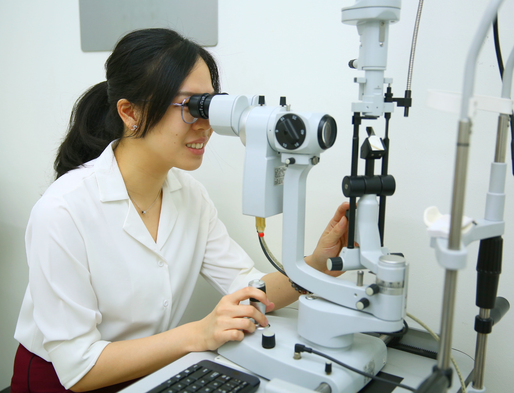
Visual Acuity Test
The visual acuity test is the fundamental part of an eye examination. It measures how well you can see at various distances. You will be asked to read letters, numbers or shapes on from a Snellen chart at a specific distance. This is the first step in identifying refractive errors such as nearsightedness, farsightedness, or astigmatism and eye related diseases.
Refraction Assessment
If the visual acuity test indicate the presence of abnormalities, our optometrist will perform a refraction assessment. During this assessment, you will undergo retinoscopy and afterwards be asked to look through a series of trial lenses while reading letters or numbers on a chart. This helps the optometrist determine the precise prescription for corrective lenses, if necessary.
Eye Muscle and Accommodation Assessment
This is where we evaluate your Binocular Vision, it includes the convergence, divergence and accommodation function. In other words it is how well your eyes work together when looking at far and near. You will be asked to follow an object with your eyes as it moves in different directions. This test helps detect any issues with eye teaming, tracking, or focusing, which can affect your overall visual performance. Further tests will follow should there be any abnormalities found.
Peripheral Vision Test
A peripheral vision test, also known as a visual field test, assesses your ability to see objects and movement outside your direct line of sight. You will be asked to focus on a central point while indicating when you see objects appearing in your peripheral vision. This test helps detect any potential blind spots or visual field defects.
Intraocular Pressure Measurement
Measuring intraocular pressure is crucial for detecting glaucoma, a group of eye condition that damages optic nerve, which includes increased pressure within the eye. Our optometrist in charge of your case may use a tonometer or a non-contact tonometer to measure the pressure. This test is painless and helps identify potential signs of glaucoma early on.
Retinal Examination
A retinal examination allows our optometrist to assess the health of your retina, optic nerve, and blood vessels at the back wall of your eye. The optometrist may use an ophthalmoscope or a fundus camera to examine the structures of your eye. This examination helps detect retinal disease & conditions such as retinal tear, retinal detachment, macular degeneration, diabetic retinopathy, and other retinal abnormalities.
Managements and Treatments
After completing all the necessary tests, our optometrist will discuss the findings and advise the management and treatment plan based on your individual needs. This may include prescribing corrective lenses, suggesting lifestyle changes, or referring you to a specialist for further evaluation, diagnosis and treatment.


